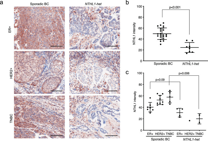
Fig. 2. NTHL1 protein expression in sporadic breast cancer (n = 21) and NTHL1-het breast cancer (n = 8).
a NTHL1 expression in sporadic breast cancer and NTHL1-het cancer of ER+, HER2+, and triple-negative types. Multiplex immunofluorescent staining approach was used and the fluorescence signal was displayed in colorimetric pattern for better contrast. NTHL1: brown color; DIPI: blue color. The epithelial marker AE1/AE3 (Supplementary Fig. 4) was used to identify cancer cells in the breast cancer tissue in NTHL1 expression quantitation in (b) and (c). Scale bar = 100 μm. b The average intensity of NTHL1 in sporadic cancer group compared to NTHL1-het group. c The average intensity of NTHL1 in sporadic cancer group compared to NTHL1-het group according to ER, PR, and HER2 status. BC breast cancer, TNBC triple-negative breast cancer.

