Figure 3.
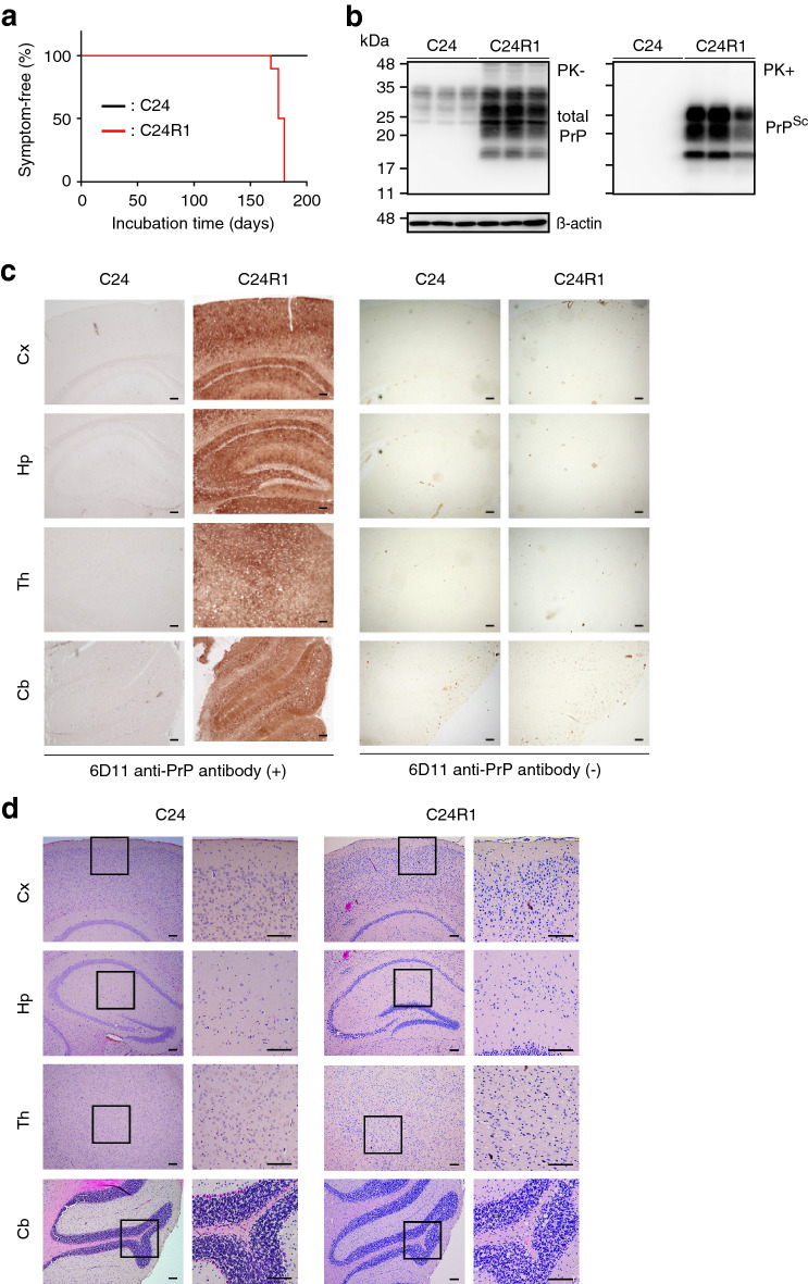
IAV/WSN infection induces de novo formation of infectious prions in N2aC24 cells. (a) The percentage of symptom-free mice after intracerebral inoculation with cell lysates from control N2aC24 and N2aC24(R1) cells. (b) Western blotting of brains from mice sacrificed at 208 dpi with the N2aC24 cell lysate and from mice symptomatic after inoculation with N2aC24(R1) cell lysate with 6D11 antibody after treatment with or without PK (20 μg/mg of proteins). PK-untreated 20 μg proteins from each brain homogenate were used for detection of total PrP and PK-treated 40 μg proteins for PrPSc. β-actin was used as an internal control. (c) Immunohistochemistry of brains from mice sacrificed at 208 dpi with the N2aC24 cell lysate and from mice terminally ill after inoculation with N2aC24(R1) cell lysate with or without 6D11 antibody. Bars, 100 μm. (d) Hematoxylin–eosin staining of brains from mice sacrificed at 208 dpi with the N2aC24 cell lysate and from mice terminally ill after inoculation with N2aC24(R1) cell lysate. Cx, cerebral cortex; Hp, hippocampus; Th, thalamus; Cb, cerebellum. Bars, 100 μm. Full length blots of Western blot images are shown in Supplementary Fig. 9a, b.

