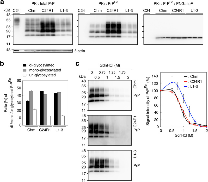
Figure 6.
Different biochemical properties of PrPSc in N2aC24(R1) cells from PrPSc in RML- and 22L-infected N2aC24 cells. (a) Western blotting of N2aC24(R1), RML-infected N2aC24Chm (Chm), and 22L-infected N2aC24L1-3 cells (L1-3) with 6D11 antibody after treatment with or without PK (20 μg/mg of proteins). PK-untreated 15 μg proteins from each cell lysate were used for detection of total PrP (Left panel) and PK-treated 30 μg proteins for PrPSc (Middle panel) Aliquots of the PK-treated samples were also treated with PNGase F before Western blotting (Right panel). β-actin was used as an internal control. (b) Ratio (%) of di-, mono-, and un-glycosylated PrPSc in the Middle panel of (a). (c) Western blotting (Left panels) of PrPSc and its signal intensities (Right panel, n = 3) in RML-infected N2aC24Chm (Chm, Upper panel), N2aC24(R1) (Middle panel), and 22L-infected N2aC24L1-3 cells (L1-3, Lower panel) with 6D11 antibody after treatment with different concentrations of GdnHCl. Full length blots of Western blot images are shown in Supplementary Fig. 11.

