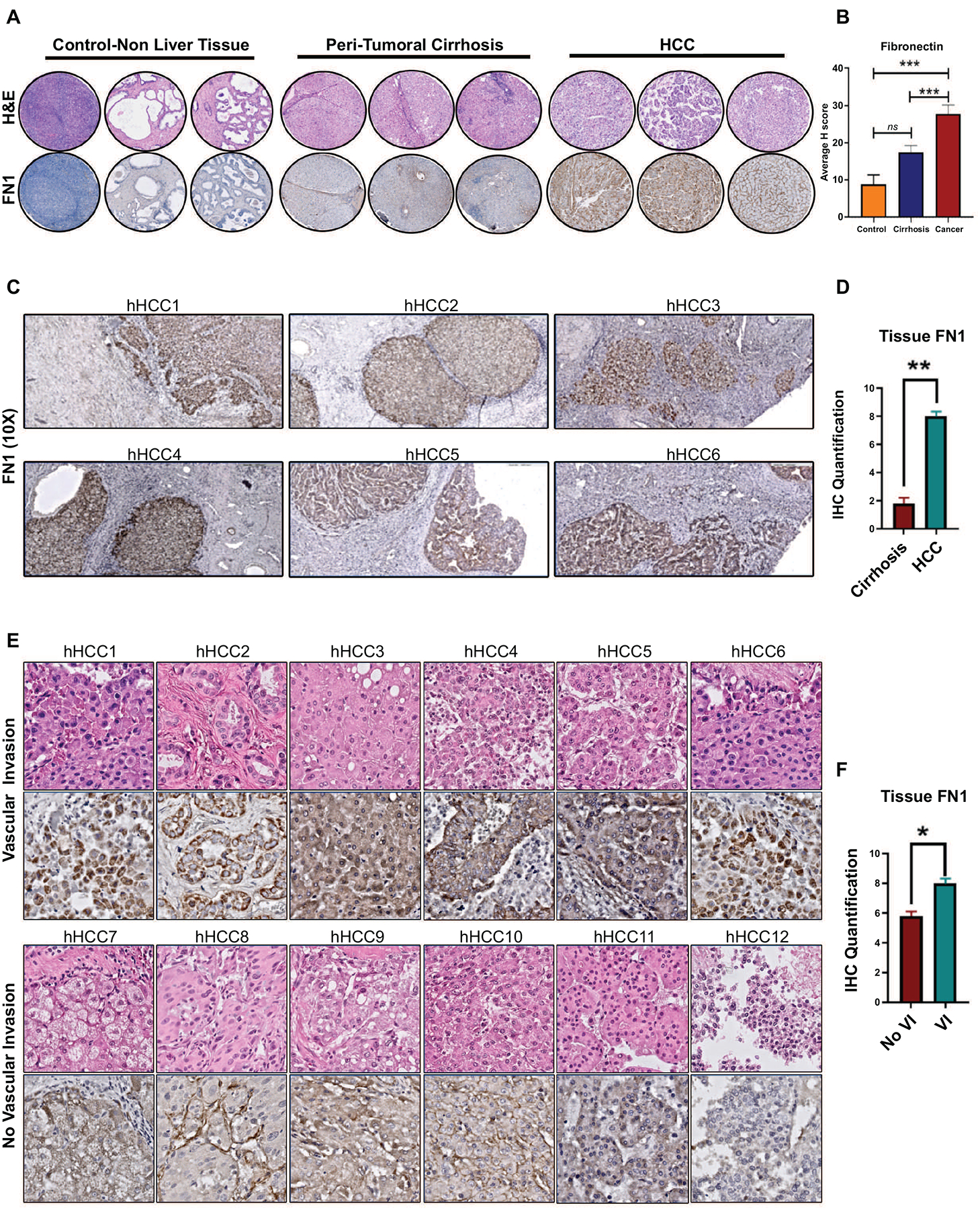
Figure 7. Validation of fibronectin tissue expression in human HCC.

a. Immunohistochemistry for fibronectin in tissue microarray - representative H&E and IHC (10x) from non-liver tissue, peritumoral cirrhosis tissue and HCC tumor tissue. b. Quantification of Fn1 expression in the tissue microarray. c. Representative immunohistochemistry for FN1 in surrounding liver and human HCC tissues with vascular invasion. d. Quantification of Fn1 expression in the whole slide tissue. e. Representative immunohistochemistry for FN1 in human HCC whole slide tissues with (top panel) and without vascular invasion (bottom panel). f. Quantification of FN1 in tumors with and without vascular invasion. * p<0.05; **p<0.01; ***p<0.001.
