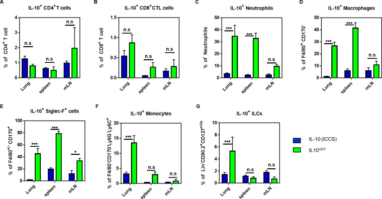
Fig 2. The myeloid-derived IL-10GFP signal is masked by autofluorescence in granulocytes.
C57BL/6 and IL-10GFP mice were challenged three times (d0, d1, d2) with either PBS or 10 μg LPS per mouse via aspiration. 16–18 hours after the last administration, the single cell suspension from lungs, spleens, and mLNs were stained for CD4+ T cells (CD45+ CD45R/B220- CD3ε+ CD4+ CD8α-), CD8+ T cells (CD45+ CD45R/B220- CD3ε+ CD4- CD8α+), macrophages (CD45+ CD45R/B220- CD3ε- Siglec-F- F4/80+), Siglec-F+ cells (eosinophils, alveolar macrophages; CD45+ CD45R/B220- CD3ε- Siglec-F+ F4/80-/+), neutrophils (CD45+ CD45R/B220- CD3ε- Siglec-F- F4/80- Ly6G+ CD11b+), monocytes (CD45+ CD45R/B220- CD3ε- Siglec-F- F4/80- Ly6G- Ly6C+ CD11b+), and ILCs (CD45+ CD45R/B220- CD3ε- Siglec-F- F4/80- Ly6G- CD90.2+ CD127lo/-). The expression of IL-10 was measured either by GFP+ or by intracellular IL-10 as indicated. (A-G) Comparison of the IL-10 signal derived from intracellular cytokine staining (ICCS) or from the GFP-signal (IL10GFP) of IL10GFP mice in (D) CD4+ T cells, (E) CD8+ T cells, (F) neutrophils, (G) macrophages, (H) Siglec-F+ cells (eosinophils, alveolar macrophages), (I) monocytes, and (J) ILCs from indicated organs. The graph shows combined data from two independent experiments (PBS: n = 6 mice/group, LPS: n = 7–9 mice/group in total).

