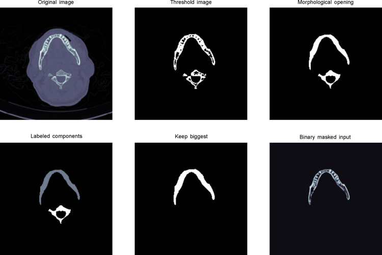
Fig 2. Mandible bone extraction from a raw slice image.
(a) Load the original image and crop its center with an image size of 256×256. (b) Apply a threshold of 400 to remove tissue and unrelated parts. (c) Perform morphological opening operation, a technique to remove small objects, while maintaining the shape and property of large objects. (d) Label components to identify objects. (e) Retain the large objects in labeled components. (f) Binary masked input to obtain the bone part of mandible slice.

