Figure 5.
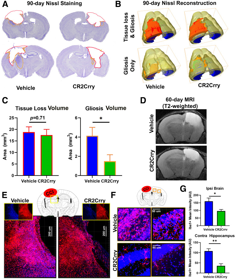
CR2Crry inhibits gliosis but not lesion volume after TBI. A, Representative Nissl immunostaining of animals treated with vehicle or CR2Crry starting day 56 (3 doses every 48 h). Brains were extracted on day 90 after TBI for analysis. Red contour represents area of tissue loss. Orange contour represents area of gliosis. B, 3D reconstruction of lesion volume from Nissl-stained serial sections (40 µm thick, 200 µm apart) using Amira to show volumes of tissue loss and gliosis. C, Quantification of volume of tissue loss and gliosis shown in B. N = 8/group. *p < 0.05 (Student's t test). D, Representative T2-weighted MRI images of animals treated as in A showing similar area of tissue loss (T2 Bright) between vehicle and CR2-Crry treated mice at 1 week after treatment. E, F, High-resolution IF staining for perilesional ipsilateral (E) and contralateral (F) cortex and hippocampus showing density of Iba1+ cells (red). G, Quantification of IF staining in E and F. N = 5 animals/group (2 or 3 sections each). *p < 0.05; **p < 0.01; Student's t test.

