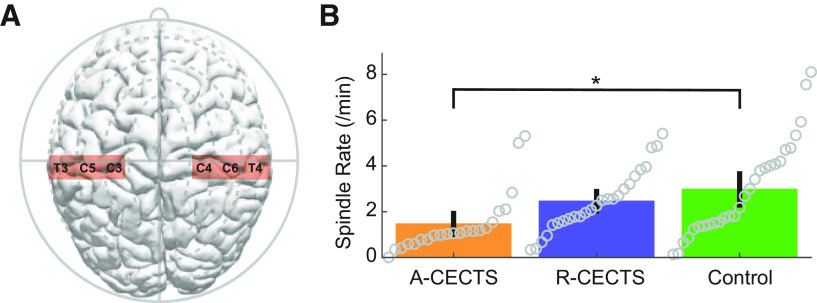
Figure 3.
Spindle rate is lower in patients with active CECTS. A, Electrodes analyzed in the centrotemporal channels of each hemisphere. B, Spindle rate versus subject group. Circles represent spindle rate for each hemisphere of each patient. Bar heights indicate the mean spindle rate of the group. Vertical black lines indicate 2 SEs of the mean. *p = 0.019.

