Figure 3.
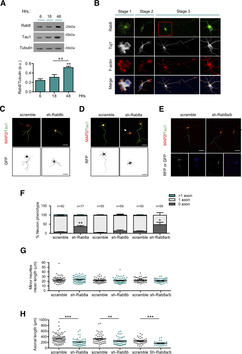
Rab8a is required for axon formation. A, Representative Western blotting and quantification of Rab8 expression from cultured cortical neurons at 6, 18, and 48 h postplating. B, Representative images of neurons at stages 1–3 stained with Rab8 (green), Tuj1 (gray), F-actin (red), and TO-PRO-3 (blue). C, Representative images of hippocampal neurons overexpressing scrambled or shRNA against Rab8b, stained with MAP2 and tau-1. D, Representative images of hippocampal neurons overexpressing scrambled or shRNA against Rab8a, stained with MAP2 and tau-1. E, Representative images of hippocampal neurons overexpressing scrambled or shRNA against Rab8a and Rab8b (1:1), stained with MAP2 and tau-1. F, Quantification of neuron phenotypes of RFP or/and GFP-positive cells in C–E. G, Quantification of mean minor neurite length in D–E. H, Quantification of axonal length in C–E. White arrow shows nucleofected neurons. Scale bars: 50 µm. Values represent mean ± SEM (n = 3); *p < 0.05, **p < 0.01, ***p < 0.001 as compared with the corresponding control; ##p < 0.01 as compared between conditions.

