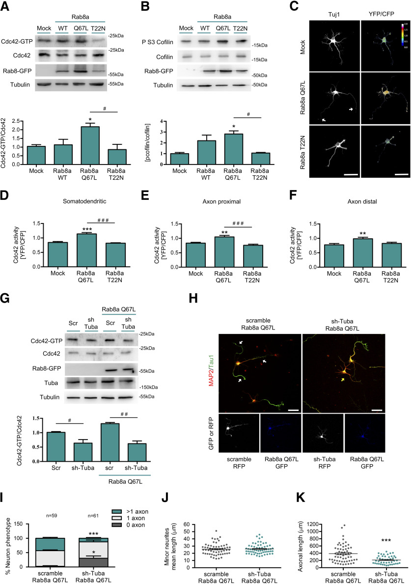
Figure 5.
Rab8a CA triggers Tuba-dependent Cdc42 activation. A, Representative image and quantification of pulldown assays from N1E-115 neuroblastoma cell lines overexpressing GFP, Rab8a WT, Rab8a Q67L (CA mutant), or Rab8a T22N (DN mutant). B, Representative image and quantification of Western blottings for cofilin and phospho ser3 cofilin from N1E-115 neuroblastoma cell lines overexpressing GFP, Rab8a WT, Rab8a Q67L (CA mutant), or Rab8a T22N (DN mutant). C, One DIV cultured hippocampal neurons expressing a FRET biosensor for Cdc42 co-expressing Flag, Flag-Rab8a Q67L, or Flag-Rab8a T22N and stained against Tuj1 (white). D–F, Quantification of the Cdc42-GTP/Cdc42 ratio from C in somatodendritic (D), proximal axonal (E), and distal axonal compartments (F). G, Representative image and quantification of pulldown assays from N1E-115 neuroblastoma cell lines knock-down for Tuba overexpressing GFP or Rab8a Q67L (CA mutant). H, Representative images of Rab8a Q67L-overexpressing neurons co-transfected with a shRNA against Tuba, stained with MAP-2 and tau-1. White arrow shows axons in multiaxonic neurons, yellow arrow shows nucleofected neurons. I, Quantification of neuron phenotypes of GFP and RFP-positive cells in H. J, Quantification of minor neurite average length in H. K, Quantification of axonal length in H. Scale bars: 50 µm. Values represent mean ± SEM (n = 3); *p < 0.05, **p < 0.01, ***p < 0.001 as compared with the corresponding control; #p < 0.05, ##p < 0.01, ###p < 0.001 as compared between conditions.

