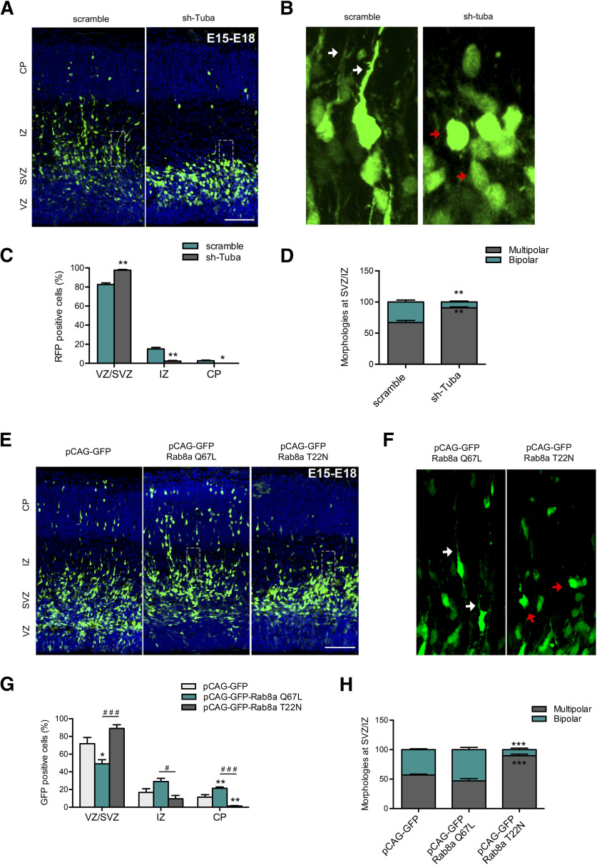
Figure 7.
Tuba and Rab8a regulates neuronal morphology of migrating neurons in the embryonic cortex. A, Representative images of cortical sections of IUE mice with pRFP-sh-scramble or pRFP-sh-Tuba, at E15 and analyzed at E18. RFP-positive cells (pseudo-colored green) were located in the CP, IZ, SVZ, and VZ. B, Representative image of a zoom between SVZ and IZ of panel A. The white arrow shows normal neurons with a bipolar morphology, while the red arrow shows cells with a round morphology. C, Quantification of the distribution of RFP-positive cells in CP, IZ, SVZ, and VZ. D, Quantification of the morphology of RFP-positive cells between IZ and SVZ. E, Representative images of cortical sections of IUE mice with pCAG-GFP, pCAG-GFP plus Rab8a Q67L, or pCAG-GFP plus Rab8a T22N, at E15 and analyzed at E18. F, Representative image of a zoom between SVZ and IZ of panel A. The white arrow shows normal neurons with a bipolar morphology, while the red arrow shows cells with a round morphology. G, Quantification of the distribution of GFP-positive cells in CP, IZ, SVZ, and VZ. H, Quantification of the morphology of GFP-positive cells between IZ and SVZ. Scale bars: 75 µm. Values represent mean ± SEM (n = 4); *p < 0.05, ***p < 0.01, ***p < 0.001 as compared with the corresponding control and #p < 0.05 and ###p < 0.001 as compared between conditions.

