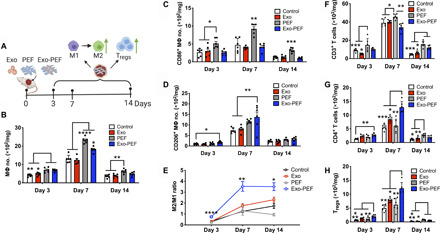
Fig. 3. Exo-PEF promoted the M2 MΦ subtype and Treg populations in the skin wound model.

(A) Full excisional skin wounds were treated with injected exosomes, PEF, or Exo-PEF. Exosome dosage: 30 μg of protein mass. Analyses of immune cell numbers per milligram of tissue (with untreated wounds as the negative control): (B) total MΦs, (C) CD86+ M1 MΦs, (D) CD206+ M2 MΦs, (E) ratio of CD206+ M2 versus CD86+ M1 MΦ, (F) total T cells, (G) CD4+ T cells, and (H) CD4+CD25+FoxP3+ Tregs on days 3, 7, and 14 after injury. n = 6. *P < 0.05, **P < 0.01, ***P < 0.001, and ****P < 0.0001.
