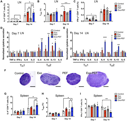
Fig. 4. Exo-PEF activated the TH2 immune response in lymphatic organs.

T cell subtypes were analyzed in lymph nodes (LN) and spleen following skin excisional wounds and treatments. (A to C and G to I) Percent analyses of IL-4+CD4+ T cells, CD4+CD25+FoxP3+ Tregs, and IFN-γ+CD4+ T cells in the inguinal LNs (A, B, and C, respectively) and spleen (G, H, and I, respectively) (n = 6; *P < 0.05, **P < 0.01, and ***P < 0.001). (D and E) Cytokine array studies on supernatants produced by ex vivo LN cells isolated from the LNs harvested on day 7 (D) and day 14 (E) after injury. (F) Representative hematoxylin and eosin (H&E)–stained images of the LNs harvested from mice (scale bar, 500 μm).
