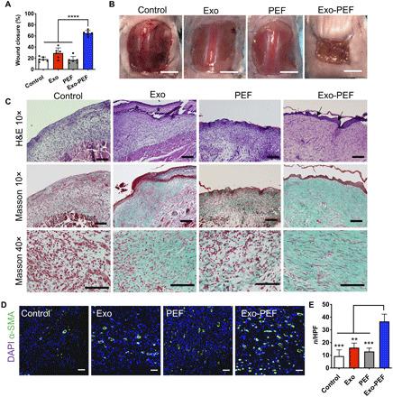
Fig. 5. Exo-PEF healed large skin wounds in 2 weeks, showing wound closure, reepithelialization, collagen deposition, and blood vessel formation.

(A) Percent wound closures and (B) representative images of the wounds after 2-week treatments. (C) H&E and Masson’s trichrome (MT) histological staining of wound tissues after 2-week treatments. Asterisks, fibrin-like granulocyte-rich layer in the Exo group; arrows, neo-formed epidermis in the Exo-PEF group. Scale bars, 200 μm (10×) and 100 μm (40×). (D) Fluorescent staining of α-smooth muscle actin (α-SMA) (green) and DAPI (blue) in the wound bed. Scale bar, 50 μm. (E) Statistical calculation of α-SMA–positive blood vessel numbers per high-power field (HPF). Exosome dosage: 50 μg of protein mass. n = 6. **P < 0.01, ***P < 0.001, and ****P < 0.0001.
