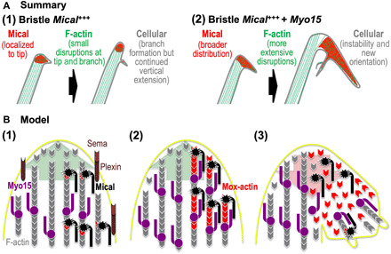
Fig. 6. Summary/model: Myo15 propagates Mical Redox-triggered F-actin disassembly and cellular remodeling.

(A) Summary: (1) Left: Mical (red) localizes to the growing bristle tip and is spatiotemporally activated to disassemble F-actin and trigger cellular remodeling/branching. Right: As development continues, the bristle again assembles F-actin/bundled filaments, which elongate the bristle past this region of F-actin disassembly/branching. (2) Left and right: Elevating Myo15 levels distributes Mical more broadly around the branch point, spatially increasing F-actin disassembly. Right: Myo15’s expansion of Mical’s distribution and F-actin disassembly hinders new F-actin/bundled filament assembly, destabilizing and misdirecting the elongating bristle. Myo15 thereby transforms regions where Sema/Plexin/Mical activation typically induces limited disassembly/reorganization (1) into more expansive effects (2). (B) Model: (1) During outgrowth/extension (large green arrow), Mical (black) specifically localizes and is activated by Sema repellents (light brown) and their Plexin receptors (dark brown) (4, 5). Activated Mical oxidizes F-actin (gray) subunits on their pointed ends, generating Mical-oxidized actin (Mox-actin) subunits (red) (6, 7, 9). Myo15 (purple) also works within these growing regions, carrying cargo toward F-actin barbed ends/the membrane (26, 41, 49). (2) Mical–Myo15 associations transport Mical and its Redox-enzymatic F-actin-modifying effects toward F-actin barbed ends/the membrane. (3) This Myo15 transport expands Mical-mediated F-actin disassembly, which breaks down specific cellular regions (large red arrow). This F-actin disassembly also derails/deposits F-actin-transported cargo specifically within these disrupted regions, which positions it to assist in the local reconstruction/branching that follows Mical-triggered F-actin disassembly.
