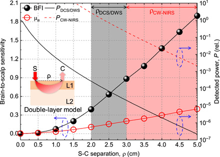
Fig. 1. Brain-to-scalp sensitivity of optical BFI (DCS/DWS) exceeds that of absorption (CW-NIRS).

The brain-to-scalp sensitivities of optical BFI and optical absorption (μa) measurements were simulated (section S5) with a double-layer model (inset), with extracerebral and cerebral layers designated as “scalp” and “brain” for short. Optical BFI is intrinsically more brain specific than absorption, achieving a higher brain-to-scalp sensitivity at a given S-C separation. However, the remitted light flux and detected power (right y axis) decrease approximately exponentially with increasing S-C separation, which is needed for high brain-to-scalp sensitivity. Because of the expense of single or few mode photon-counting channels, required for coherence, DCS/DWS uses relatively short S-C separations (black shading). On the other hand, CW-NIRS can collect many modes that sum incoherently and therefore can use larger S-C separations (red shading). We assume that PCW − NIRS is 104 × PDCS/DWS, based on a hypothetical CW-NIRS system that collects 104 modes for every single DCS/DWS mode. Blue dashed ovals with arrows point to corresponding y axes. Inset: ρ (S-C separation), L1 (extracerebral layer), and L2 (cerebral layer). Note that in practice, effective brain-to-scalp sensitivity can be further improved by considering additional superficial short S-C channels in signal analysis.
