. 2021 Apr 19;10:e61769. doi: 10.7554/eLife.61769

Figure 5. Netrin 1 (NTN1) and deleted in colorectal carcinoma (DCC) regulate midline zipper glia (MZG) organization during interhemispheric fissure (IHF) remodelling.

(A) Gfap-positive mature astroglia (green or white in inset), Glast-positive glia (red) and pan-Laminin (LAM)-positive IHF and basement membrane (magenta) in embryonic day (E)17 wildtype Dcckanga, Dcc knockout and Ntn1-LacZ mice. Yellow arrowheads indicate presence (filled) or absence (open) of midline glial populations, the MZG, indusium griseum glia (IGG) and glial wedge (GW). Fluorescence intensity of Gfap staining from insets or bins in insets (red dotted line) was quantified in (B) and (C). (D) Schema of MZG development, IHF remodelling and corpus callosum (CC) formation in wildtype mice and mice deficient for NTN1 or DCC. Red dotted lines indicate rostral and caudal bins that were used to calculate the ratio of GFAP fluorescence in (C). All graphs represent mean ± SEM. Statistics by Kruskal–Wallis test with post-hoc Dunn’s multiple comparison test. ***p<0.001; ns: not significant. See related Figure 4—figure supplement 1, Figure 5—figure supplement 1 and Supplementary file 1.

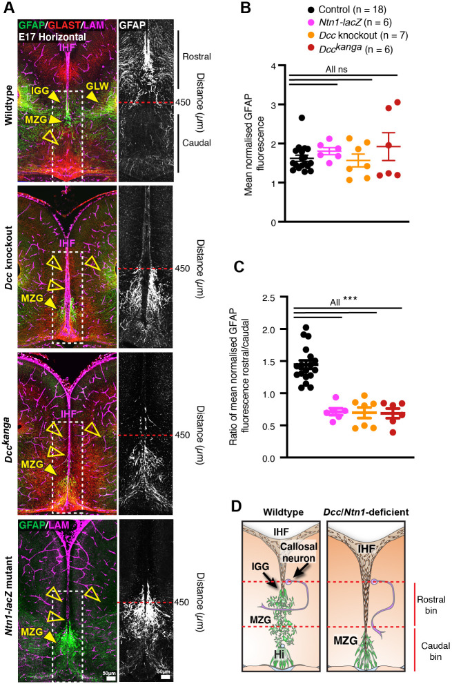
Figure 5—source data 1. Normalized fluorescence intensity of GFAP adjacent to the telencephalic midline in E17 Dcc and Ntn1 mutant mice.
elife-61769-fig5-data1.xlsx (799.7KB, xlsx)

Figure 5.

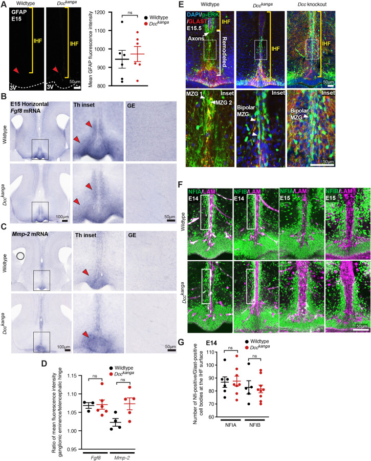
Figure 5—figure supplement 1. Deleted in colorectal carcinoma (DCC) is not required for astroglial differentiation of midline zipper glia (MZG).

Figure 5—figure supplement 1.

(A) Mature astroglial marker Gfap (white) in horizontal sections of embryonic day (E)15 wildtype and Dcckanga mice with quantification of Gfap average fluorescence intensity. The surface of the third ventricle (3V) is outlined with dotted white lines. Red arrowheads indicate reactive blood vessels that are not Gfap-positive glia. Yellow brackets indicate the position of the interhemispheric fissure (IHF). Fgf8 mRNA (B) or Mmp-2 mRNA (C) in horizontal sections of E15 wildtype and Dcckanga mice. Red arrowheads indicate reactivity in the telencephalic hinge (Th) in insets, right. A region of the ganglionic eminence (GE) where Fgf8 is not expressed is shown and was used to normalize specific Fgf8 expression within the Th with background immunoreactivity as quantified in (D). (E) Phosphorylated p44/42 Mapk or Erk1/2 (p-ERK, green) and Glast-positive MZG (red) in horizontal sections of E15.5 wildtype, Dcckanga mice and Dcc knockout mice reveal the extent of IHF (yellow brackets) and remodelled regions of the septum (white brackets) with p-ERK-positive MZG in insets. (F) Nuclear factor I (NFI) A or B (green) and pan-Laminin (LAM)-positive leptomeninges and basement membrane (magenta) in horizontal sections of E14 and E15 wildtype and Dcckanga mice. NFI-positive/Glast-positive MZG cell bodies at the IHF surface are outlined with white boxes and quantified in (G). Data is represented as mean ± SEM. Significant differences were determined with nonparametric Mann–Whitney tests. ns: not significant.
Figure 5—figure supplement 1—source data 1. Fluorescence intensity measurements for GFAP, Fgf8 and Mmp-2 mRNA, and quantification of nuclear factor I (NFI)-positive/GLAST-positive cell bodies in Dcckanga mice.