Figure 1.
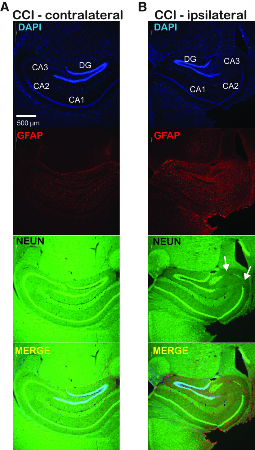
CCI produces persistent astrogliosis and loss of CA3 neurons that are restricted to the ipsilateral hippocampus. A, Immunofluorescent images of contralateral hippocampus one month after CCI with DAPI (blue; nuclear), GFAP (red; astrocyte), NEUN (green; neuron), and merged images. Hippocampal subregions are labeled within the DAPI panel. B, Immunofluorescent images from ipsilateral hippocampus after CCI showing DAPI, GFAP, NEUN, and merged images as in A. Persistent astrogliosis after CCI is seen as increased GFAP staining, arrows indicate region of neuronal loss in the CA3 region within the NEUN panel. The astrogliosis and loss of CA3 neurons seen in ipsilateral hippocampus one month after CCI is absent in in the contralateral hippocampus; there is relative preservation of DGGCs targeted for our recordings, indicating these cells remain viable after CCI. Scale bar (500 μm) in upper left panel applies to all images.

