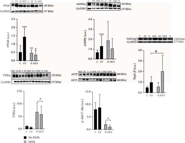
Figure 2.

Plot and western blot images showed the expression of proteins in the heart forty-eight hours after injection with doxorubicin (DOX). Groups: C: control; GT: green tea; D: doxorubicin; DGT: doxorubicin+green tea. PPARα: peroxisome proliferator-activated receptor; AMPKα: activated protein kinase; TNFα: tumor necrosis factor-alpha; P-akt/T-akt: protein kinase B phosphorylated; Top2-β: topoisomerase IIβ. Pi: P value for interaction between factors GT and DOX. Pgt: P value for the differences between the groups with GT and without GT; Pd: P value for the differences between the groups with DOX and without DOX. For PPARα: Pi = 0,001 and AMPKα: Pi = 0, 04, there were interactions between GT and DOX. Thus, for PPARα and AMPKα, we considered ∗DGT ≠ C, ∗∗D ≠ GT, ∗∗∗C ≠ GT, and ∗∗∗∗D ≠ GT. Comparisons without interaction considered the groups #DOX (D + DGT) ≠ no DOX (C + GT); > (GT + DGT) ≠ (C + D). For TNFα, Pd = 0, 01 and for P-akt/T-akt, Pd = 0, 02. For Top2-β, Pgt = 0, 03.
