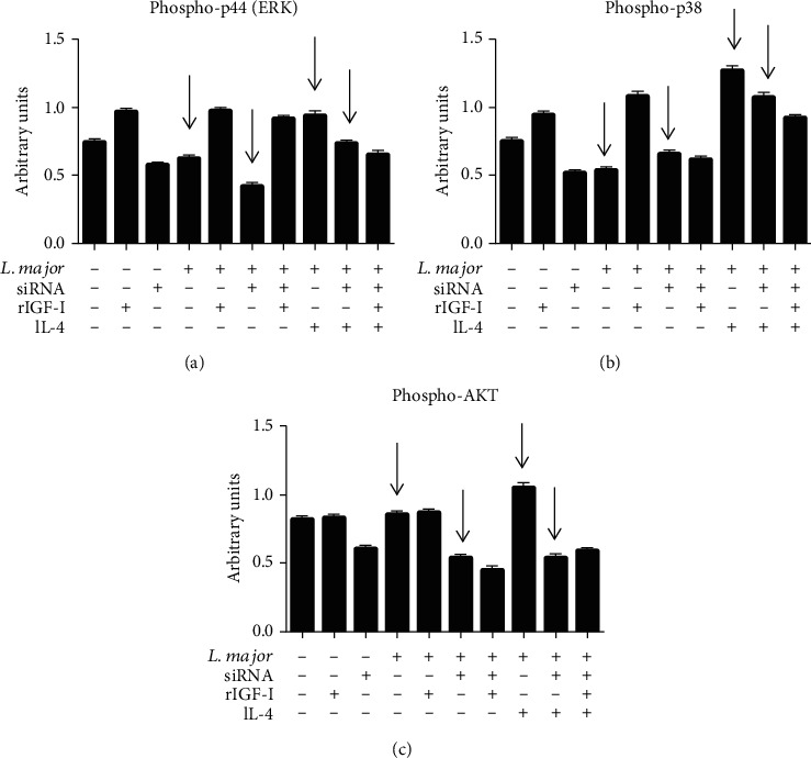
Figure 6.

The effects of IGF-I siRNA and IL-4 on components of the IGF-I signaling pathways: levels of phosphorylated p44 (ERK), p38 (MAPK), and AKT proteins. L. major promastigote-infected or noninfected RAW 264.7 cells transfected with or without IGF-I siRNA were stimulated for 30 minutes with IL-4 (2 ng/mL; R&D Systems, EUA) and recombinant IGF-I (50 ng/mL; R&D Systems, EUA). Cells were lysed, the proteins were separated in 10% SDS-PAGE, and subsequently, a Western blotting was performed using anti-phospho-p44 (137F5, Cell Signaling Technology, USA), anti-phospho-p38 (D13E1, Cell Signaling Technology, USA), and anti-phospho-AKT (Ser473, Cell Signaling Technology, USA) antibodies. Protein bands corresponding to protein expression levels were submitted to a densitometric analysis (AlphaEaseFC™ software 3.2 beta version; Alpha Innotech Corporation, USA), and data are expressed in arbitrary units (adapted from Reis et al. [65]).
