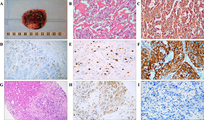
Fig. 1.
Key gross, microscopic and immunohistochemical findings of PPGLs. A Macroscopic appearance of the resected adrenal with a 10 cm encapsulated pheochromocytoma exhibiting a fleshy cut surface with solid tan-colored areas. B Photomicrograph of hematoxylin–eosin (H&E)-stained tumor tissue at × 400 magnification revealing a nested growth pattern and an exceedingly well-vascularized stroma. C Chromogranin A immunostaining at × 400 magnification. Note the diffusely positive cytosolic staining. D Sustentacular cells visualized using an S100 staining. E The Ki-67 proliferation index can be used to assess the proliferative activity, and is also a key part of certain algorithms to assess the metastatic potential. F Positive SDHB immunohistochemistry argues against underlying SDHB, C, and D gene mutations, thereby indicating a lower risk of disseminated disease. G Core needle biopsy of a liver metastasis in a patient previously resected for a pheochromocytoma 2 years earlier, with metastatic tumor cells recognizable through their basophilic cytoplasm. These tumor cells were positive for neuroendocrine markers (not shown). Upper left portion depicts hepatocytes. H GATA3 immunohistochemistry displaying nuclear positivity. I Complete loss of sustentacular cells was noted, as evident by an S100 immunostaining. This phenomenon is often reported in metastatic cases

