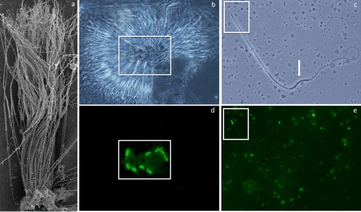
Figure 1.
Whitespotted bamboo shark Chiloscyllium plagiosum spermatozuegmata and spermatozoa micrographs illustrating the morphology and acrosome location for aggregated and individual spermatozoa. Scanning electron micrograph of a spermatozeugma fragment (a) revealing sperm with heads closely aligned and embedded in a matrix core. Matching phase contrast and fluorescent micrographs of a spermatozeugma with radially aligned heads (b, d) and individual spermatozoan (c, e) with acrosomes highlighted by fluorescein isothiocyanate conjugated Arachis hypogaea agglutinin (PNA-FITC) stain. Whitespotted bamboo shark spermatozoa (c) possess a helical head with an acrosome (e), a helical midpiece and a flagellum with a transient cytoplasmic sleeve (arrow) at the junction of the midpiece and tail that was shed after acquiring motility.

