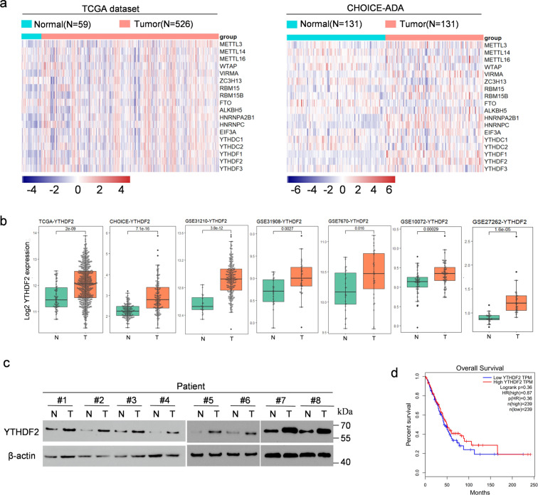
Fig. 1. YTHDF2 was highly expressed in LUAD.
a Heat map profiling the expression of m6A WERs in TCGA (left) and CHOICE (right) databases of LUAD. b Relative RNA levels of YTHDF2 in LUAD tissue and normal lung tissue in TCGA, CHOICE, and GEO datasets. c Immunoblotting assay of YTHDF2 expression in eight paired LUAD primary tumor samples. β-actin was used as a loading control. d) Kaplan–Meier analysis of LUAD cancer in TCGA for the correlations between YTHDF2 expression and overall survival.

