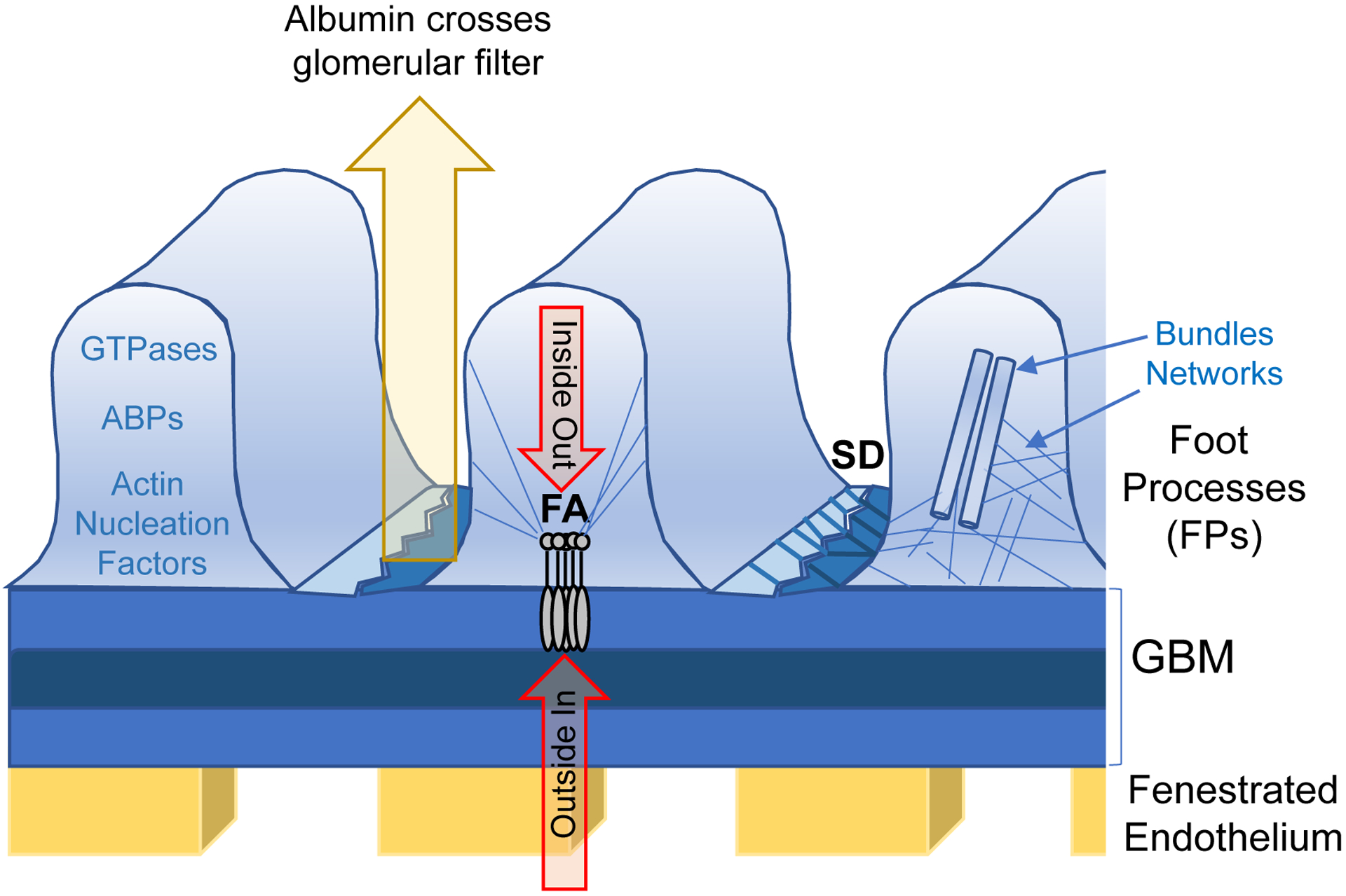
Figure 1. Schematics of the glomerular filter.

Specificity of the kidney filter is maintained by fenestrated endothelium, the glomerular basement membrane (GBM) and glomerular podocytes. The structure of podocyte foot processes (FPs) is regulated by two signaling platforms: focal adhesions (FAs) and the slit-diaphragm (SD). Those two hubs integrate ‘inside out’ and ‘outside in’ signaling, which defines the global organization of the actin cytoskeleton. The status of the actin cytoskeleton is defined by regulatory GTPases, actin binding proteins (ABPs) and actin nucleation factors. Two major actin structures are tight actin bundles and loose networks, both of which are established by actin crosslinking proteins.
