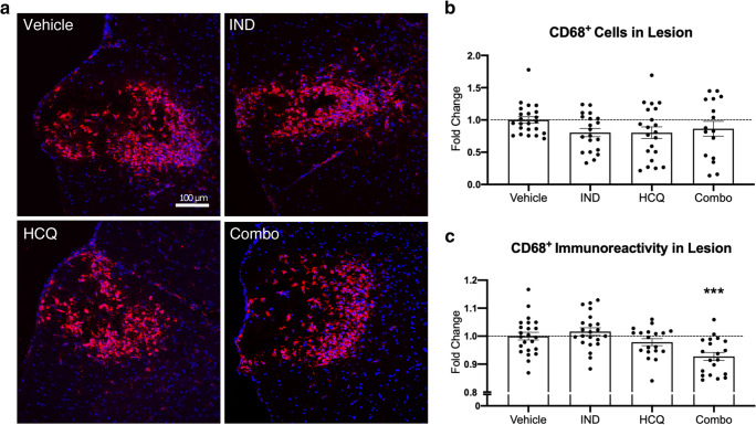
Fig. 6.
Effect of drug treatments on CD68+ microglia/macrophages in vivo. (A) Representative images of the day 3 lysolecithin lesion, visualizing DAPI for nuclei (blue) and CD68 for microglia/macrophages within the lesion (red). (B) Number of CD68+ cells within the lesion, expressed as percent coverage of the lesion area. (C) Mean fluorescence intensity of CD68+ microglia/macrophages within the lesion. Lower immunoreactivity represents reduced cell activation. Pooled data from the three separate experiments (n ≥ 5 each), normalized and represented as fold change of the vehicle group. White dashed line depicts lesion area. Graphs represented are the mean with SEM. Data were analyzed using a one-way ANOVA with Dunnett’s post hoc multiple comparison test, compared to vehicle treatment. Each data point represents one animal. Horizontal dashed line represents fold change of 1 (the mean of vehicle group). ***p < 0.001 compared to vehicle control. IND: indapamide, HCQ: hydroxychloroquine

