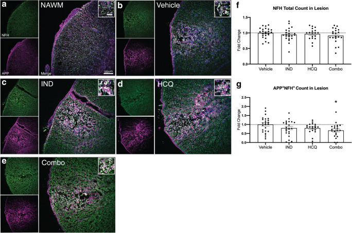
Fig. 7.
Combination treatment reduces the magnitude of axonal damage in vivo. (A–E) Representative images of normal-appearing white matter (NAWM), or of lysolecithin lesions in mice treated with vehicle, IND, HCQ, and combination. Each image includes NFH for axons (green), APP for compromised axons (magenta), and an overlay image. The insets of the overlay image depict APP accumulation on NFH+ axons (white). (F) Total number of NFH+ axons in the lesion. (G) Quantitation of number of NFH+ axons with accumulated levels of APP, represented as fold change of vehicle group. Graphs represent the mean with error bars depicting SEM. Data were analyzed using a one-way ANOVA with Dunnett’s post hoc multiple comparison test; *p < 0.05 compared to vehicle treatment. Each data point represents one animal. Pooled data from the three separate experiments (n ≥ 5 each), normalized and represented as fold change of the vehicle group. Horizontal dashed line represents fold change of 1 (the mean of vehicle group). IND: indapamide, HCQ: hydroxychloroquine

