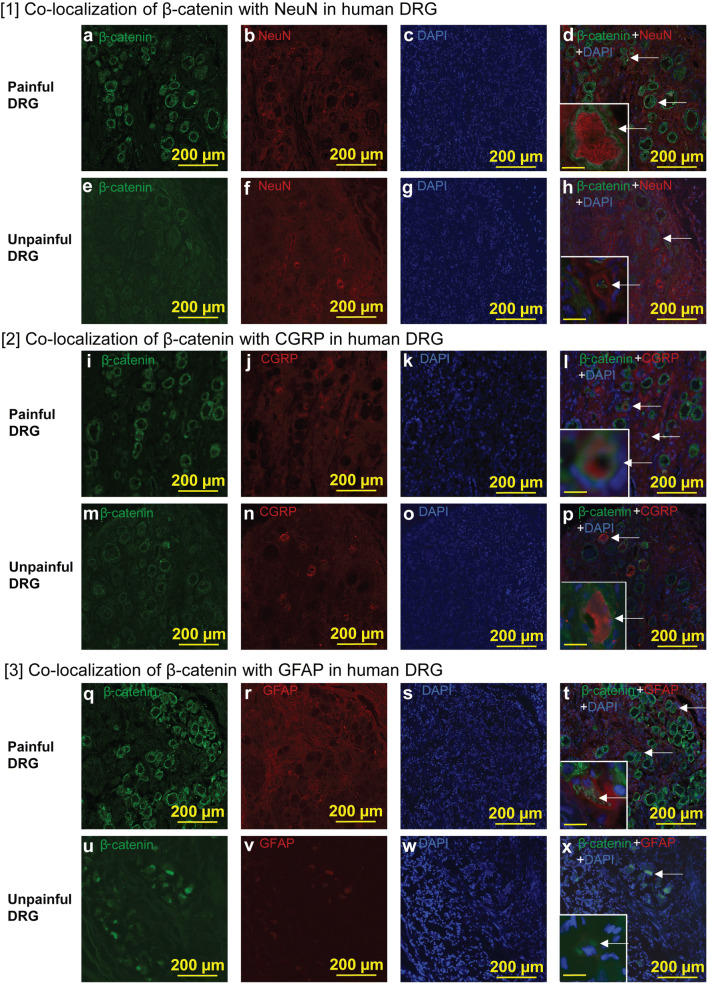
Fig. 5.
Co-localization of β-catenin in DRG neurons and satellite cells in human DRG. (1) Co-localization of β-catenin with NeuN in human DRG. (a) The DRG was isolated from the pain area of a patient. Note that the DRG neurons from this area had increased expression of β-catenin (green) as a canonical Wnt signaling marker. (b) NeuN (red) in the DRG from the pain area. (c) DAPI (blue) in the DRG from the pain area. (d) β-Catenin (green), NeuN (red), and DAPI (blue) in the DRG from the pain area. β-Catenin was expressed in neurons. Arrows indicate β-catenin with NeuN. Scale bar in the inset was 20 μm. (e) DRG was isolated from outside the pain area of a patient. Note that the DRG neurons from outside the pain area expressed low levels β-catenin (green) compared with that in the pain area (a). (f) NeuN (red) in the DRG from outside the pain area. (g) DAPI (blue) in the DRG from outside the pain area. (h) β-Catenin (green), NeuN (red), and DAPI (blue) in the DRG from outside the pain area. Scale bar in the inset was 20 μm. (2) Co-localization of β-catenin with CGRP in human DRG. (i) The DRG was isolated from the pain area of a patient. Note that the DRG neurons from the pain area had increased expression of β-catenin (green) compared with that in the unpainful area (m). (j) CGRP (red) in the DRG from the pain area. (k) DAPI (blue) in the DRG from the pain area. (l) β-Catenin (green), CGRP (red), and DAPI (blue) in the DRG from the pain area. Arrows indicate β-catenin with CGRP. β-Catenin was expressed in CGRP-expressing neurons. Scale bar in the inset was 20 μm. (m) The DRG was isolated from outside the pain area of a patient. β-Catenin (green) in the DRG from outside the pain area. (n) CGRP (red) in the DRG from outside the pain area. (o) DAPI (blue) in the DRG from outside the pain area. (p) β-Catenin (green), CGRP (red), and DAPI (blue) in the DRG from outside the pain area. Arrows indicate β-catenin with CGRP. Scale bar in the inset was 20 μm. (3) Co-localization of β-catenin with GFAP in human DRG. (q) DRG was isolated from the pain area of a patient. Note that the DRG neurons from the pain area had increased expression of β-catenin (green) compared with that in the unpainful area (u). (r) The GFAP (red) in the DRG from the pain area. (s) DAPI (blue) in the DRG from the pain area. (t) β-Catenin (green), GFAP (red), and DAPI (blue) in the DRG from the pain area. Arrows indicate β-catenin with GFAP. β-Catenin was expressed in GFAP-expressing satellite cells. Scale bar in the inset was 20 μm. (u) The DRG was isolated from outside the pain area of a patient. β-Catenin (green) in the DRG from outside the pain area. (v) GFAP (red) in the DRG from outside the pain area. (w) DAPI (blue) in the DRG from outside the pain area. (x) β-Catenin (green), GFAP (red), and DAPI (blue) in the DRG from outside the pain area. Arrows indicate β-catenin with GFAP. Scale bar in the inset is 20 μm. Scale bars, 200 μm

