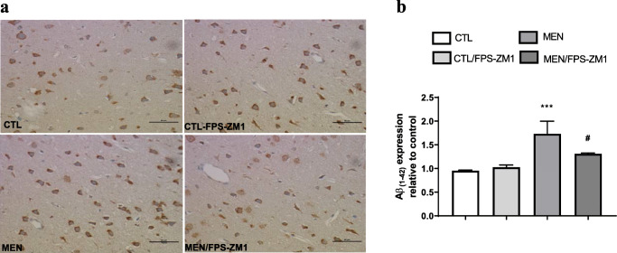
Fig. 13.
Immunohistochemical staining of Aβ1–42 in rat brain prefrontal cortex. (a) Representative microscopic field images (magnification × 400) immunostained with Aβ1–42. (b) Data are presented as the mean ± SEM (n = 3-4). ***p < 0.001 as compared to the control group. #p < 0.05 as compared to the meningitis group

