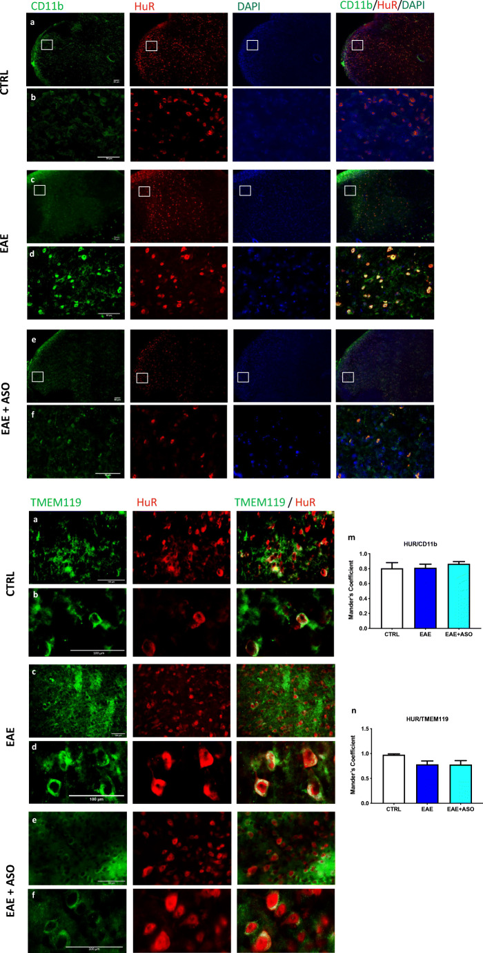
Fig. 2.
Localization of HuR protein in activated microglia from EAE spinal cord. Fluorescence microscopy images showed the microglial localization of HuR in the spinal cord of control (CTRL), EAE, or EAE + anti-HuR ASO mice. a, c, e: Low-magnification images show a homogeneous distribution of HuR within the spinal cord. Immunostaining for the microglial marker CD11b in the same section reveals similar immunoreactivity. The merged image shows HuR localization on microglial cells. b, d, f: High-magnification images show the cytosolic localization of HuR, consistent with a pattern of activation, and merged images illustrate the high degree of colocalization of HuR and CD11b. Nuclei were counterstained with DAPI. Antibodies are shown at the top. Scale bar = 50 μm. Expression of HuR protein on TMEM119-positive microglial cells. Fluorescence microscopy images showed the microglial localization of HuR in the spinal cord of control (CTRL), EAE, or EAE + anti-HuR ASO mice. g, i, k: low-magnification images. h, j, l: high-magnification images. Antibodies are shown at the top. Evaluation of colocalization of HuR and CD11 (m) or TMEM119 (n) in spinal cord sections. Scale bars = 100 μm. CTRL, dODN-treated nonimmunized mice; EAE, dODN-treated EAE mice; EAE + ASO, anti-HuR ASO-treated EAE mice

