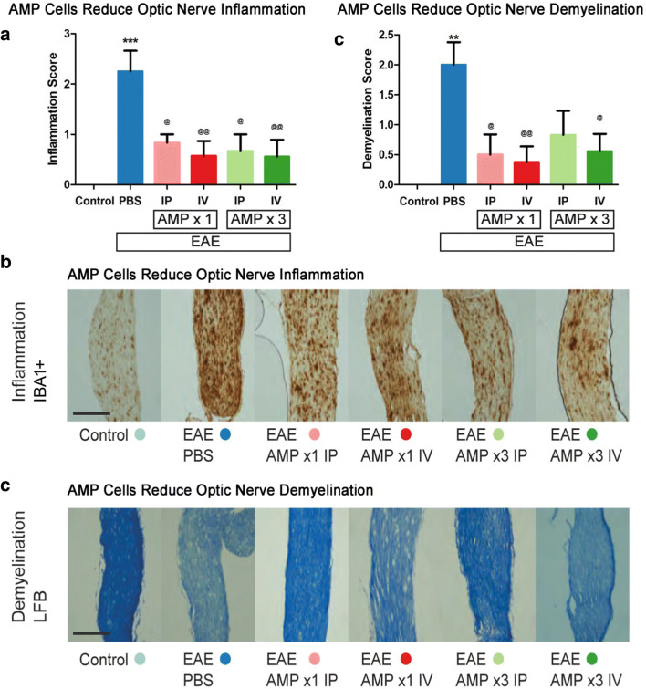
Fig. 3.
AMP cells suppress optic nerve inflammation and demyelination. a Immunomodulatory effects of systemic AMP cells in the optic nerve was evaluated by IBA1 staining and scored using a 4 point scale. Vehicle (PBS) treated EAE mice showed a significant (***p < 0.05) increase in optic nerve (N = 8) inflammation as compared with control (N = 6) mouse optic nerves. All AMP cell treatment paradigms (1× IP, N = 6; 1× IV, N = 7; 3× IP, N = 6; 3× IV, N = 9) significantly decreased inflammation in the optic nerves as compared with vehicle-treated EAE mice (@p < 0.05; @@p < 0.01). b Images show IBA1+ cells in one representative optic nerve section (original magnification × 10, Scale bars: 200 μM). Few microglia/macrophages are observed in the control mouse optic nerve, whereas the vehicle-treated mouse optic nerve contains numerous cells and optic nerves from AMP cell-treated EAE mice contain only moderate IBA1+ cell levels. c Demyelination in the optic nerve was evaluated by LFB staining and scored using a 3 point scale. Vehicle-treated EAE mice showed a significant (**p < 0.01) increase in optic nerve demyelination as compared with control mouse optic nerves. AMP cell treatment with 1× IP and IV injection, and 3× IV injection, significantly decreased demyelination in the optic nerves as compared with vehicle-treated EAE mice (@p < 0.05; @@p < 0.01), and 3× IP injection of AMP cells lead to a non-significant trend in attenuation of myelin loss. d Representative images of optic nerves stained with LFB show a vehicle-treated EAE mouse with myelin loss in the optic nerve, AMP cell–treated mouse optic nerves with partial preservation of myelin. Original magnification × 20, Scale bars: 200 μM. Data represent mean ± SEM of measurements from the right eye of each mouse

