Fig. 3.
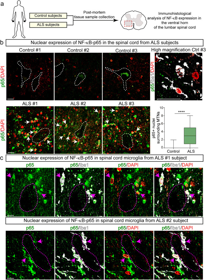
Nuclear NF-κB-p65 increased expression in microglia surrounding motor neurons in autopsied ventral spinal cords from ALS subjects. (a) Scheme of the experimental design. b) Representative confocal images showing the expression of nuclear NF-κB-p65 in the cellular microenvironment surrounding spinal motor neurons (white dotted lines) of ALS and control subjects. Upper panels: NF-κB-p65 staining (green) and DAPI (red) in control subjects. Note the lack of nuclear NF-κB-p65 expression. The panel to the right represents a higher magnification image of few Iba1-positive microglia (white) that lack cytoplasmatic or nuclear expression of NF-κB-p65 (green). Lower panels: represent confocal images showing a systematic increase expression of nuclear NF-κB-p65 in the surroundings of motor neurons in 3 sporadic ALS subjects. Arrows indicate yellow nuclei with NF-κB-p65 colocalizing with DAPI. The graph to the right shows the quantitative analysis of NF-κB-p65-positive nuclei surrounding motor neurons. All quantitative data are expressed as mean ± SEM; data were analyzed by the Mann-Whitney test, ****p < 0.0001. n = 3 ALS patients and 3 controls. Scale bars = 20 μm. (c) Higher magnification confocal images showing the cytoplasmic and nuclear expression (magenta arrows) of NF-κB-p65 in Iba1-positive microglia in the ventral spinal cord of ALS subjects. Scale bars = 10 μm

