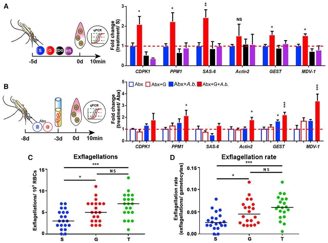
Figure 6. The increase of pH induces male gametogenesis.

(A) Quantification of the mRNA abundance of genes associated with male gametogenesis in mosquitoes fed with S, G, 2-DG, and HS.
(B) Quantification of the mRNA abundance of genes associated with male gametogenesis in A. bogorensis re-colonized mosquitoes fed with S and G.
(C and D) Exflagellations and exflagellation rate in the mosquitoes fed on S, G, and T. Exflagellations were monitored 10 min post-infection. The median number of exflagellations per 105 erythrocytes is shown (C), and exflagellation rate was shown as the percent (exflagellations/gametocytes) per 105 erythrocytes (D).
Significance in (A) and (B) was determined by ANOVA followed by Dunnett’s tests. Error bars indicate SEM (n = 5). Each dot represents one mosquito, and horizontal lines represent the medians in (C) and (D). Data shown in (C) and (D) were pooled from two independent experiments. Significance was determined by ANOVA followed by Holm Sidak’s tests. NS, not significant, *p < 0.05, **p < 0.01, ***p < 0.001. S, 2% sucrose; G, 2% sucrose + 0.1 M glucose; T, 2% sucrose + 0.1 M trehalose; 2-DG, 2% sucrose + 0.1 M glucose + 5 mM 2-DG; HS, 10% sucrose. See also Figure S5.
