Fig. 1—

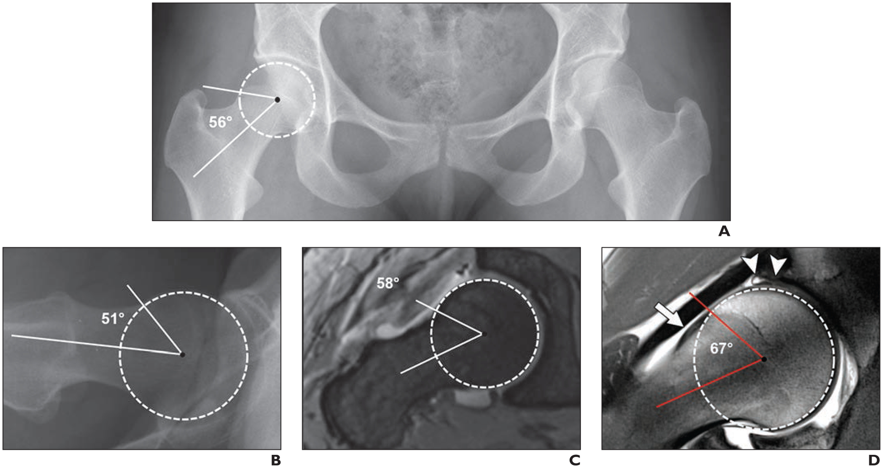
28-year-old woman with 2-year history of right-sided groin pain and positive anterior impingement test (pain with hip flexion, adduction, and internal rotation). Case emphasizes importance of appropriate imaging workup of patients with suspected femoroacetabular impingement to arrive at correct diagnosis. Alpha angles are measured between femoral neck axis (bottom line) and line that connects proximal part of asphericity (top line), determined by best-fitting circle (circle), with femoral rotation center.
A, Anteroposterior radiograph of pelvis shows normal pelvic morphology, preserved hip joint space, and no arthritic change.
B, Cross-table lateral radiograph of symptomatic right hip shows no obvious deformity. Patient was referred for MR arthrography to evaluate morphology of femoral head-neck junction.
C, Oblique axial MR arthrogram shows normal femoral head-neck junction.
D, Radial proton density–weighted MR image shows mild cam deformity (arrow) with alpha angle of 67°. Intrasubstance tear of superior labrum with adjacent cartilage damage (arrowheads) is evident. Patient was referred for hip arthroscopy for cam resection and labral repair. Cam deformities are frequently located in anterolateral aspect of femoral head-neck junction and therefore are not visible on standard anteroposterior and cross-table lateral radiographs.
