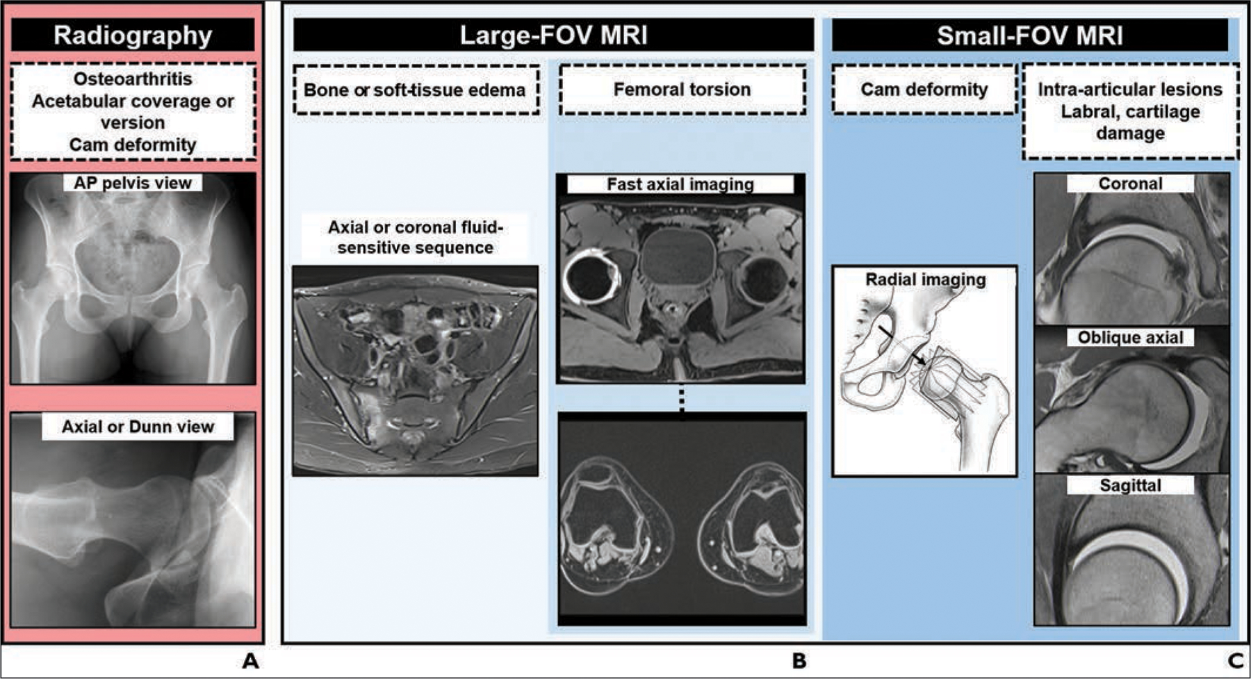
Fig. 7—

Charts show algorithm for imaging workup of patients with femoroacetabular impingement.
A, Radiography. AP = anteroposterior.
B, Large-FOV MRI.
C, Small-FOV MRI. (Drawing adapted with permission of Wolters Kluwer Health, Inc. from Steppacher SD, Tannast M, Werlen S, Siebenrock KA, Femoral morphology differs between deficient and excessive acetabular coverage, Clinical Orthopaedics and Related Research, 466, 4, 782–790, journals.lww.com/clinorthop/, a publication of The Association of Bone and Joint Surgeons)
