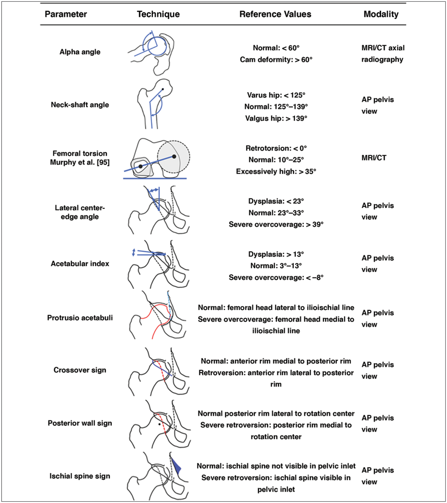
Fig. 8—

Chart shows imaging findings and reference values in femoroacetabular impingement. Alpha angle is measured between femoral neck axis (bottom line) and line that connects proximal part of asphericity (top line), determined by best-fitting circle, with femoral rotation center. Neck-shaft angle is measured between femoral neck axis (top line) and femoral shaft axis (bottom line). Femoral torsion is measured between top line connecting femoral head center, determined by perfectly fitting circle, to center of femoral neck base directly superior to lesser trochanter and bottom line connecting posterior contours of proximal femoral condyles. Lateral center-edge angle is measured between left line connecting femoral rotation center to lateral extension of sclerotic rim and vertical reference line. Acetabular wall lines shown are anterior rim (solid black lines) and posterior rim (dashed lines). Angle shown for acetabular index is formed between top line connecting medial and lateral sclerotic margins and horizontal reference line. For protrusio acetabuli, femoral head contour (red line) overlaps with ilioischial line (blue line). In crossover sign, anterior rim (blue line) projects laterally to posterior rim (red line). In posterior wall sign, posterior rim (red line) projects medially to rotation center. In ischial spine sign, ischial spine (blue shading) is visible in pelvic inlet. References values are recommendations and may differ by institutional approach [22, 23, 58]. AP = anteroposterior. (Translated by permission from Springer Nature Customer Service Centre GmbH: Springer Nature Radiologe [Impingement of the hip], Schmaranzer F, Hanke M, Lerch T, Steppacher S, Siebenrock K, Tannast M, Copyright 2016)
