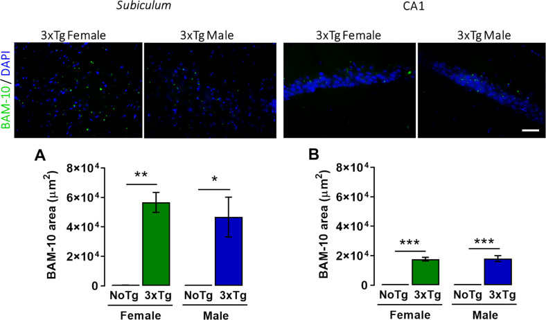
FIGURE 4.
Representative images of β-amyloid (green) immunohistochemistry and nuclei detection (blue) obtained with 40× objective in the subiculum and CA1 of the dorsal hippocampus of female and male 3xTg-AD at 5 months old. Mean BAM-10 area (μm2) (with standard error) of female and male non-transgenic (NoTg) or Alzheimer’s disease triple-transgenic (3xTg-AD) mice in the (A) subiculum and (B) CA1. *p < 0.05, **p < 0.001, ***p < 0.0001; n = 4 mice per group. Scale bar = 20 μm.

