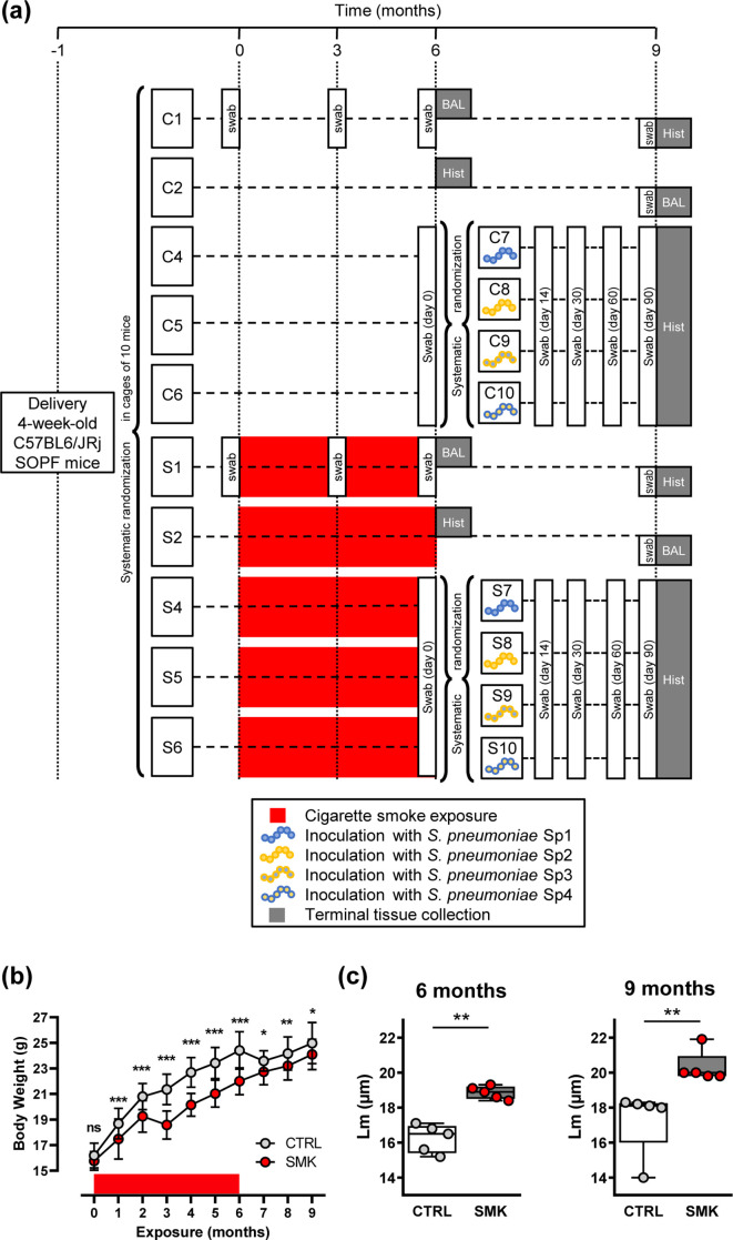
Fig. 1.
Cigarette smoke induces irreversible emphysema. (a) Experimental scheme showing randomization of purchased female mice into cages of 5–10 mice. After a 4 week acclimation period, the experiment was initiated (time point 0). Groups of mice were exposed to cigarette smoke (cages S1, S2, S4, S5 and S6) or to room air (cages C1, C2, C4, C5, C6) for 6 months. Red boxes represent exposure to cigarette smoke. Note that smoke exposure was stopped at the 6 month time point for all groups. After 6 months of exposure, three sub-groups of control mice (C4, C5 and C6) and smoke-exposed mice (S4, S5 and S6) were further systematically randomized into four groups per exposure regimen and inoculated with 2×10⁶ c.f.u. S. pneumoniae . The different strains were Sp1 (106.66 wild-type, serotype 6B), Sp2 (208.41 wild-type, serotype 7F), Sp3 (106.66 capsule mutant cps208.41, serotype 7F) and Sp4 (208.41 capsule mutant cps106.66, serotype 6B). The times of sampling of the microbiota with oropharyngeal swabs are shown. Analysis of lung structure (Hist) or BAL was performed in sub-groups of CTRL and SMK mice that were euthanized at 6 and 9 month time points. (b) Body weight was measured monthly. Data are shown as mean±sd (n=50 CTRL; n=55 SMK) and were analysed by two-way ANOVA (*** P<0.001, ** P <0.01, * P <0.05, ns (not significant). (c) Structural analysis of the lung parenchyma was performed at the indicated time points. Scatterplots show data for individual mice; median and interquartile range (box) are shown. Data were analysed by Mann–Whitney U test (** P <0.01).

