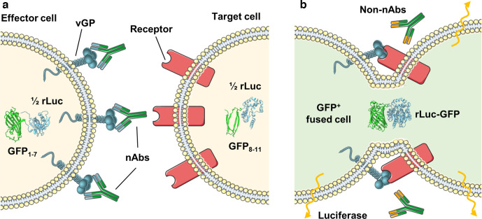
Fig. 1.
The micro-fusion inhibition test (mFIT). Samples containing antibodies are incubated with effector cells (HEK293T Lenti rLuc-GFP 1–7) expressing the viral glycoprotein (vGP) of interest. The antibody–effector cell mix is then co-cultured with target cells (HEK293T Lenti rLuc-GFP 8–11) expressing the corresponding vGP’s cellular receptor and incubated for 18–24 h. In (a) the presence of fusion-inhibitory neutralizing antibodies (nAbs) prevents the reconstitution of the rLuc-GFP reporter in fused cells, while in (b) the absence of specific neutralizing antibodies (non-nAbs), allows vGP-mediated cell–cell fusion to occur. Subsequent mixing of the target and effector cell cytoplasm leads to reconstitution of the split reporter and increased GFP and luciferase signals. This figure was generated using modified images from SMART Servier Medical Art By Servier, used under CC BY 3.0, https://smart.servier.com/, accessed June 2020.

