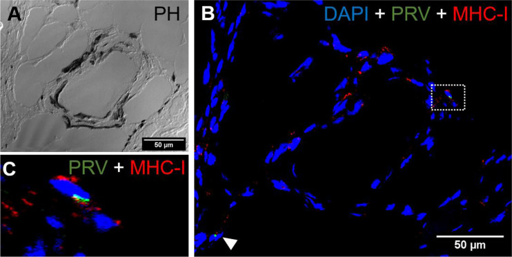
Figure 10.
Fluorescent in situ hybridization of PRV-1 and MHC-I in black focal changes. (A) phase contrast image showing vacuolar area surrounded by melano-macrophages and other immune cells. (B) Merged image showing presence of numerous MHC-I positive cells (red) where some are co-staining with PRV-1 (green) (dotted rectangle and arrowhead). (C) Magnified area from image B showing co-localization of PRV-1 in some MHC-I cells. Scale Bar = 50 µm.

