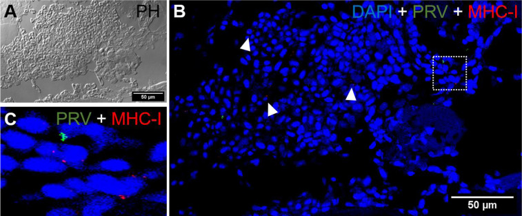
Figure 8.
Fluorescent in situ hybridization of PRV-1, and MHC-I in areas of red focal changes. (A) Phase contrast image showing a large hemorrhage. (B) Merged image of PRV-1 and MHC-I showed no co-expression of PRV-1 in MHC-I cells, but a few cells were detected in the bleeding area (arrowhead). (C) Magnified image from image B (dotted rectangle) showing PRV-1 infected cells along with MHC-I cells. Scale Bar = 50 µm.

