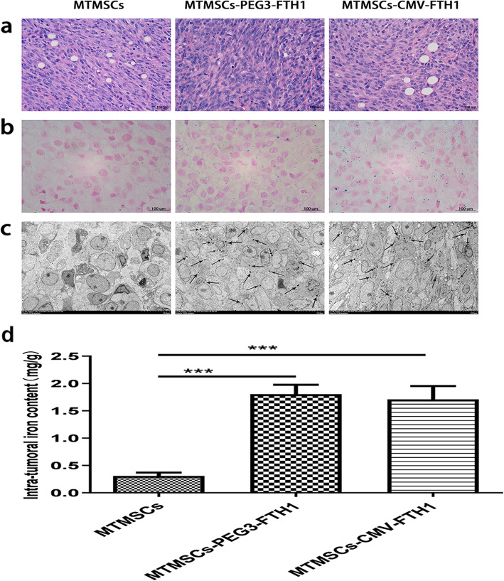
Fig. 8.
Pathological characteristics of xenografts. HE staining showed that the cells in the mass were arranged close together and that the cytoplasmic ratio was relatively large (a), which conformed to the morphology of tumor tissue. Prussian blue staining (b) and TEM (c) showed a large amount of iron particles in the mass tissue in the MTMSC-PEG3-FTH1 and MTMSC-CMV-FTH1 groups but not in the MTMSC group. The intratumoral iron content (d) was not significantly different between the MTMSC-PEG3-FTH1 and MTMSC-CMV-FTH1 groups but was significantly higher in both of these groups than in the MTMSC group (***P<0.001)

