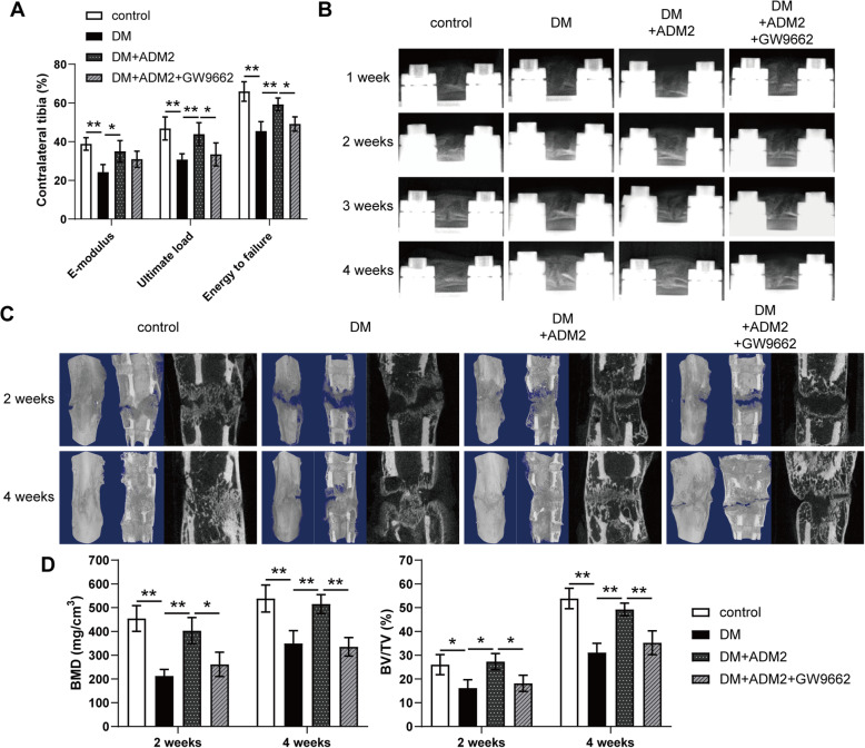
Fig. 4.
ADM2 accelerated bone consolidation during distraction osteogenesis in diabetic rats. A Mechanical tests (E-modulus, ultimate load, and energy to failure) of the distracted tibias. The values were normalized to the corresponding contralateral normal tibias. B Representative X-rays of distraction regenerate at various time points of the consolidation phase in the four groups. C Representative 3D and longitudinal images of the tibial distraction zone after 2 and 4 weeks of consolidation and treatment. D Quantitative analysis of bone mineral density (BMD) and bone volume/tissue volume (BV/TV) of the regenerated bone. The data were confirmed by one-way analysis of variance (ANOVA) followed by Tukey’s post hoc test from three independently repeated tests and are presented as the means ± SD. *P < 0.05; **P < 0.01

