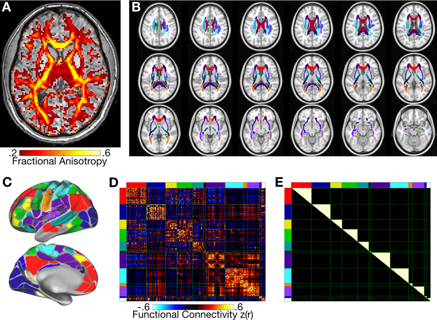
Figure 2: Data collected in the present study.

A: Diffusion Tensor Imaging scans were collected in order to produce maps of Fractional Anisotropy (FA), a measure of white matter integrity. An FA map is shown for one example subject. B: FA was assessed in each of many different a priori white matter tracts. Each tract is shown in a different color. C: Resting-state fMRI scans were collected, and data were averaged within each of 333 a priori parcels on the cortex. D: Temporal correlations were computed between all parcel timecourses to produce a functional connectivity value between each pair of parcels. A matrix illustrating the strength of functional connectivity between each parcel pair is shown for a single subject. The matrix is ordered by the known network organization of this parcel set (compare colored blocks on axes to parcel colors in C). E: For each subject, functional connectivity values from D were averaged across all unique within-network connections, as illustrated here in white.
