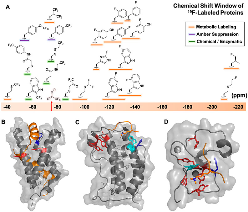
Figure 1.
(A) Chemical shift ranges of fluorine-labels incorporated in proteins, referenced to trifluoroacetate (red). Representative proteins (gray) and ligands (orange) for PrOF NMR in (B) KIX with MLL and c-Myb (PDB: 2AGH), (C) BRD4-D1 with acetylated histone H4 (PDB: 3QZT), and (D) PHD finger of BPTF with trimethylated histone H3 (PDB: 2F6J). Tyrosines (red), tryptophans (cyan), and phenylalanines (blue) within 10 Å of binding sites.

