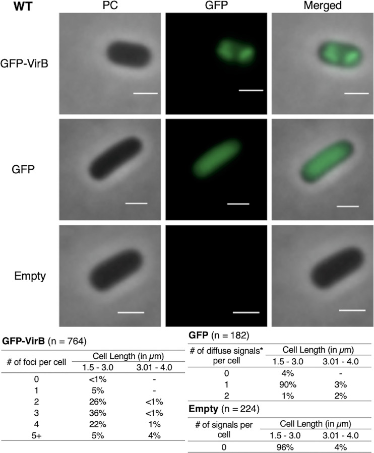
FIG 3.
Live-cell imaging of GFP-VirB in a wild-type strain of S. flexneri and quantification of foci observed during live-cell imaging of GFP-VirB in wild-type S. flexneri using MicrobeJ. Representative cells are shown. Phase contrast (left column), fluorescence (middle column) (GFP row, 44.5-ms exposure; GFP-VirB and empty, 219-ms exposure), and merged (right column) images of GFP-VirB, GFP, and an empty plasmid control . Bars, 1 μm. Within tables, a hyphen indicates that no cells fell into this category in any of the images captured; *, maxima detected by MicrobeJ.

