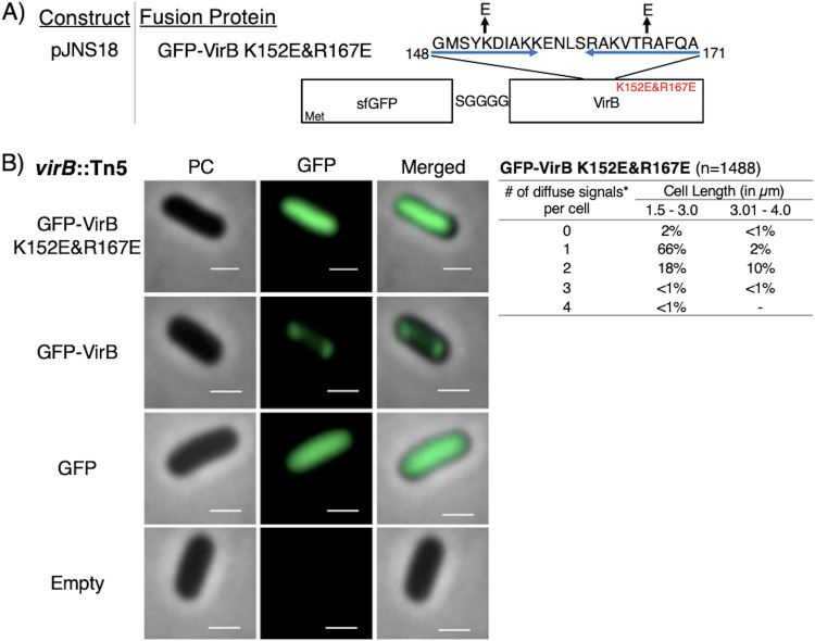
FIG 5.
Construction of a GFP-VirB DNA-binding mutant and live-cell imaging of this fusion in a virB mutant strain of S. flexneri. (A) Construct producing GFP-VirB with two amino acid substitutions, K152E and R167E, in the helix-turn-helix DNA-binding domain (denoted with blue arrows; adapted from reference 52). (B) Quantification of fluorescent signals observed during live-cell imaging of GFP-VirBK152E/R167E in virB mutant S. flexneri using MicrobeJ. Note that diffuse signals observed for this fusion were detected as maxima by MicrobeJ. Representative cells are shown. Phase-contrast (left column), fluorescence (middle column) (GFP row, 77-ms exposure; GFP-VirB, GFP-VirB K152E/R167E, and empty rows, 348.8-ms exposure), and merged (right column). Bars, 1 μm. Within the table, a hyphen indicates that no cells fell into this category in any of the images captured; *, maxima detected by MicrobeJ.

