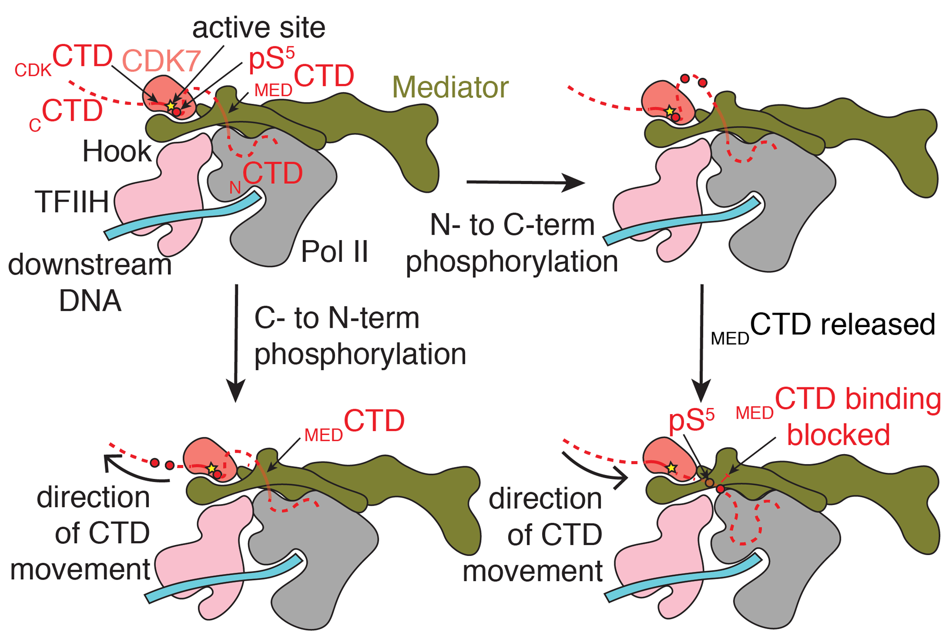
Fig. 6.

Model for phosphorylation of the Pol II CTD by CDK7. MEDCTD binding positions the rest of the CTD in the CDK7 active site. Following phosphorylation, indicated by a red circle, translocation of the CTD towards the N-terminus (bottom) would place phosphorylated repeats further from the nascent RNA emerging from Pol II. Separation of Mediator and Pol II would be difficult without separation of the CAK module and Mediator. Translocation of the CTD towards the C-terminus would position phosphorylated repeats to block binding of the CTD at MEDCTD, a possible way to favor disassembly of Med-PIC. Phosphorylated repeats would also be significantly closer to the RNA exit tunnel of Pol II to recruit the capping complex properly. CAK = cyclin-activated kinase module; CTD = C-terminal domain of RPB1; pS5 = phosphorylated serine 5 residue (red circle).
