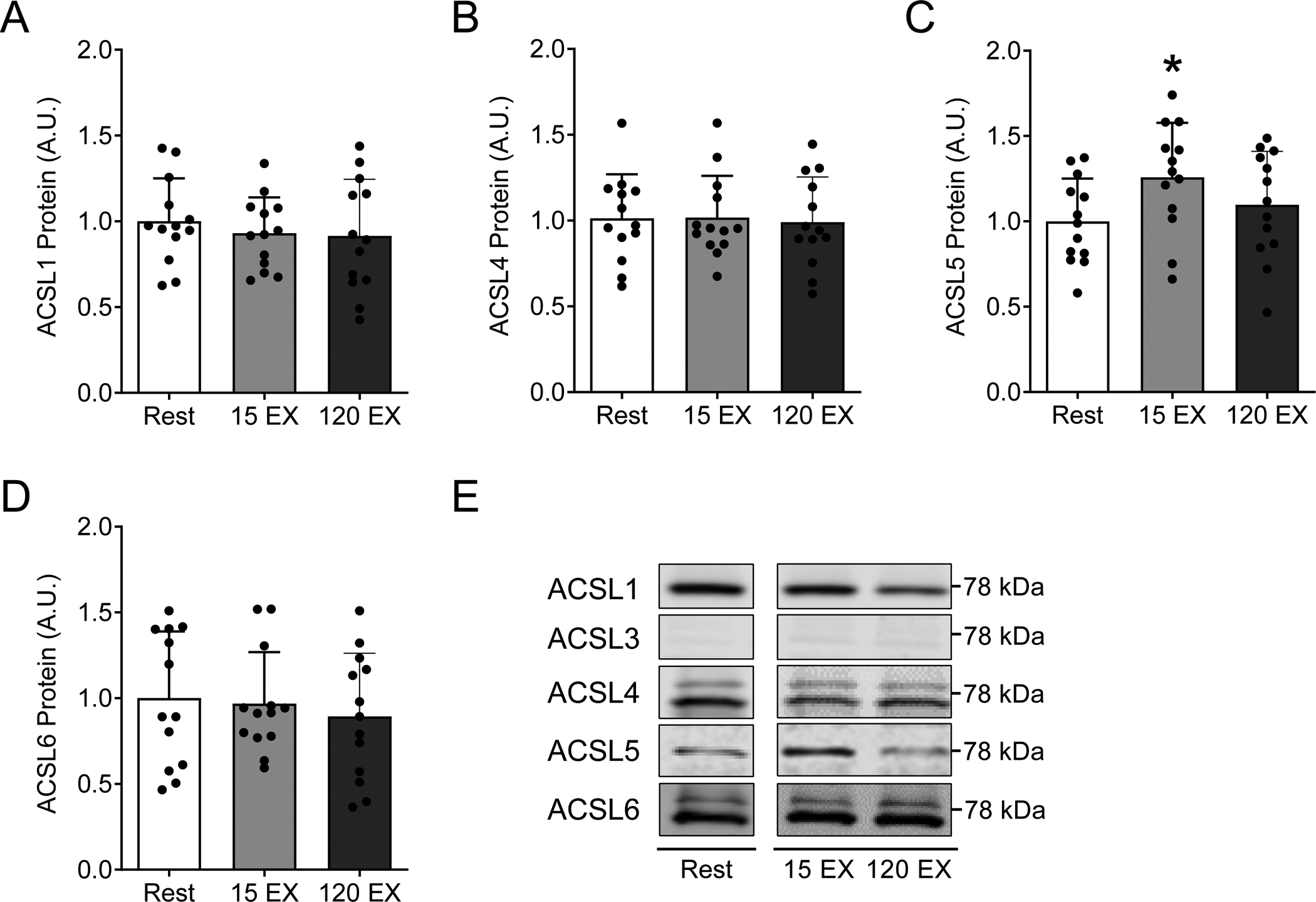
Figure 2: Skeletal muscle ACSL isoform protein abundance at rest and following acute exercise.

ACSL isoform protein abundance in vastus lateralis muscle biopsy samples at rest, 15 minutes after acute exercise (15 EX), and 120 minutes after acute exercise (120 EX). Total protein abundance for A) ACSL1, B) ACSL4, C) ACSL5, and D) ACSL6, with E) representative western blot images. Each representative image was spliced from the same blot/image for the purpose of highlighting samples reported in this study, with no alterations to the images. Full blot and ponceau images for each representative image were made available during the review process. The effects of acute exercise on skeletal muscle ACSL protein abundance were analyzed by repeated measures one-way analysis of variance models, with Dunnett’s posthoc analysis comparing post exercise time-points to rest. Data are presented as mean and standard deviation with individual data points shown. *P≤ 0.05 vs. Rest. n=14 (female/male: 10/4).
