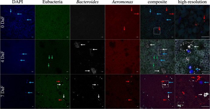
FIG 7.
False-color FISH micrograph of Macrobdella decora ILF. From left to right, columns contain images obtained with the probes DAPI (blue, eukaryotic DNA), Eub338 (green, Eubacteria), CF319a (white, Bacteroidetes [Bacteroides]), and Aer66 (red, Aeromonas) and composite and high-resolution composite (enlarged areas are indicated with white squares in the composites) images. From top to bottom, the rows contain images from animals sacrificed 0 days (wild-caught animals), 4 days, and 7 days after a laboratory-administered sterile blood meal (DaF). Blue arrows indicate eukaryotic cells (most likely leech hemocytes), green arrows indicate notable bacteria not labeled by the two specific probes, white arrows indicate Bacteroidetes microcolonies, and red arrows indicate Aeromonas. Background fluorescence is from crop contents, especially the blood meal at 4 and 7 DaF. Few bacteria are present in the crop of wild-caught, unfed animals. Bacteroidetes colony expansion occurs by 4 DaF, while Aeromonas prevalence increases by 7 DaF. Other bacteria are present at 4 DaF; however, their numbers appear to be overwhelmed by Bacteroidetes and Aeromonas growth at 7 DaF. In animals at 7 DaF, Aeromonas organisms are occasionally found associated with hemocytes. Bars = 10 μm.

