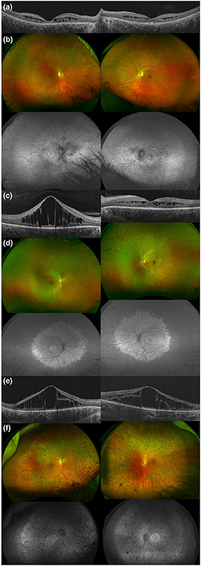
FIGURE 1.
Ophthalmologic characteristics of patients with mild peroxisomal biogenesis disorders. (a, c, and e) Macular optical coherence tomography images of Patients A, B, and C, respectively, showing cystic spaces and discontinuity of the outer retinal layers. (b and d) Color and autofluorescence ultrawide-field retinal images of patients with Heimler syndrome (Patients A and B). Their retinal dystrophy is characterized by few pigment redistribution, mild vessel attenuation, and a mostly mid-peripheral involvement with a hyper-hypoautofluorescence pattern. (f) Color and autofluorescence ultrawide-field retinal images of Patient C. In this case, we notice pigment clumping throughout the retina, moderate–severe vessel thinning, and a more extensive hyper-hypoautofluorescence (“salt and pepper”) pattern

